
Friedrich-Alexander-Universität Erlangen
Lehrstuhl für Mustererkennung
Martensstraße 3
91058 Erlangen
As a member of the Medical Image Processing group I develop automatic and semi-automatic algorithms to detect eye abnormalities and diseases. My research focuses mainly on segmentation methods. The inputs are common or spectral filtered eye fundus photographs, and fundus video sequences. Check the following two images for a sample of common, and spectral filtered fundus photographs.
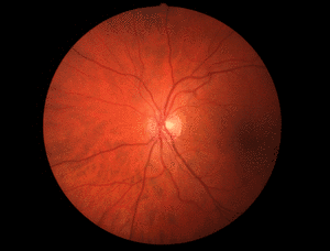 | 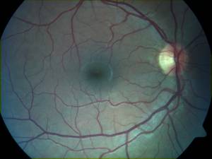 |
Red channel usually oversaturated | The color channels are balanced |
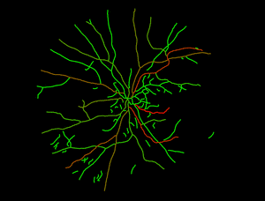
The vessel tree and other tissues like optic disc and the macula region will be detected and analysed to extract information about physioligy and diseases of the eye. The following two example show the extracted tortuosity information of the vessels and the optical density of macular pigments.
 | 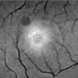 |
of macular pigments |