Daniel Stromer M. Sc.
Researcher in the Learning Approaches for Medical Big Data Analysis (LAMBDA) group at the Pattern Recognition Lab of the Friedrich-Alexander-Universität Erlangen-Nürnberg
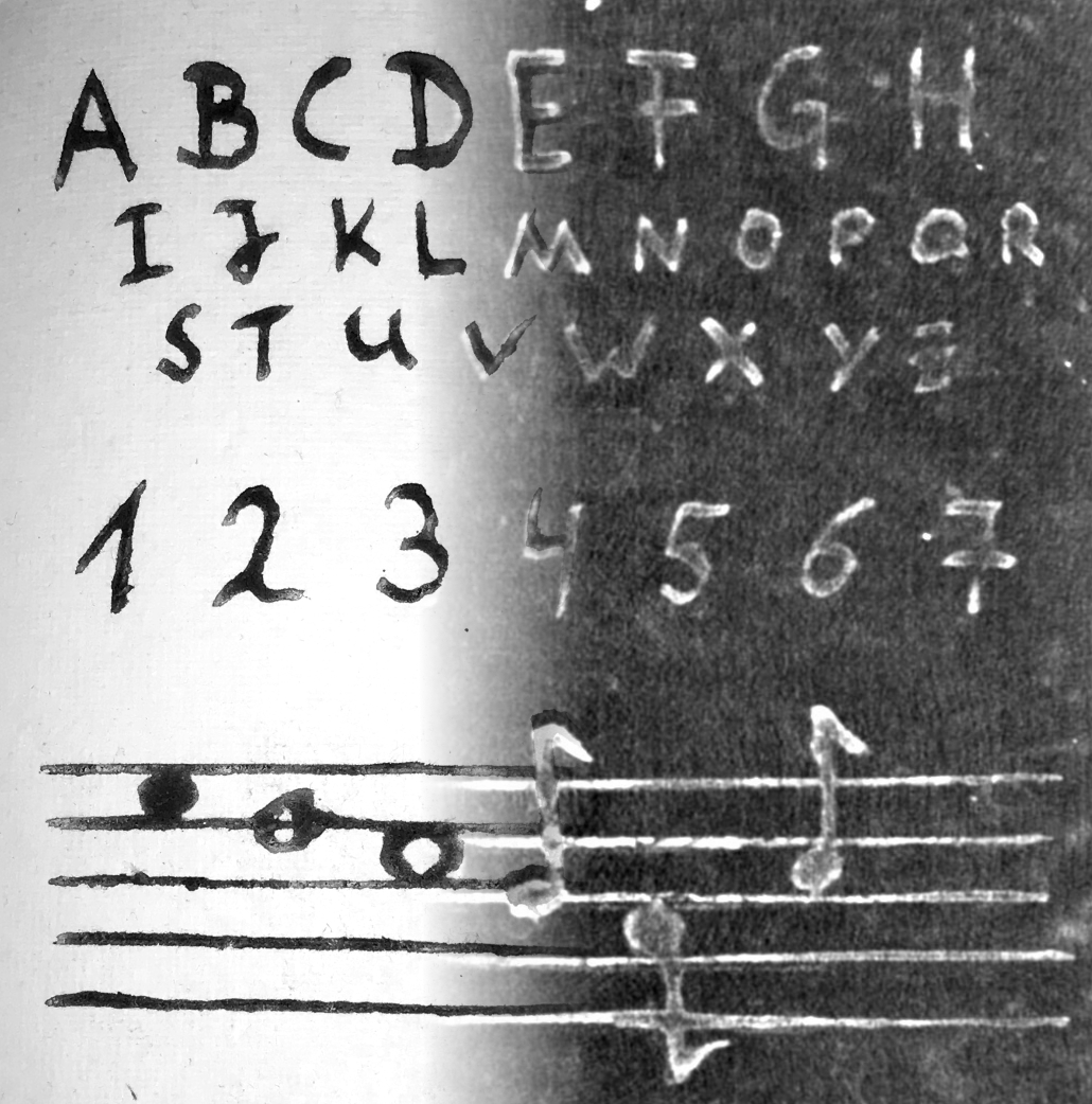
Documents are relics of the past that give us insights into long forgotten times. In former times, those hand-crafted documents were highly diverse depending on the time and culture when they were produced. From papyri scrolls in Egypt, Bamboo scrolls in Asia, paper-based codices covered in ivory and gold to the digital formats that we use today. The current trend is to digitize all those old documents ('massive digitization') and use data-driven methods to find connections and gather new insights to the past that are not obvious when investigating each document on its own ( The standard digitization method is to use a scan robot that automatically browses the pages and captures images. However, there are documents that are too fragile to open them such that the content would be damaged. Non-invasive imaging methods are capable of taking a look inside such objects. In this project, we reveal the hidden writings by means of conventional 3D X-ray computed tomography and present complete pipelines - from scanning to image processing and data compression. We create realistic scenarios and show the possibilities as well as the limitations of the method for European and Asian manuscripts. European Books: Chinese Bamboo Scrolls: |  |
Detailed description at: ![]() click here
click here
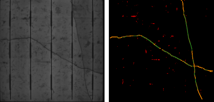
The annually produced quantity of solar modules has steadily increased over the past decades. To monitor the production outcome of such cells, errors have to be detected in a non-invasive manner. To localize cracks in solar cells, luminescence imaging is used, where several approaches for an automatized inspection exist, but a standard solution for an automatized inspection algorithm is not yet available. This is, in particular, true for multicrystalline solar cells, where the grainy structures in the luminescence images are hard to distinguish from small cracks. This project aimed to propose a segmentation algorithm for cracks and to reduce the number of false-positives while keeping the true-positives high. The image shows an EL image of a solar cell with cracks (left) and the algorithms result (right).

The connective tissue between the fat layer and the skin termed fascia has been of interest to the clinical and zoological research to study normal skin echogenicity, thickness and hydration status, as well as the echogenicity patterns of various pathological conditions. The current state-of-the-art method for visualizing these layers is to use ultrasound imaging. By visual inspection of those (Figure below), one can see four different layers: skin, fat, fascia and muscle. Our goal is to separate the different layers fully automatically by applying appropriate segmentation algorithms. Furthermore, we want to provide a GUI for specialist such that there is no more need to manually measure the layers.


 +49 9131 85 25246
 +49 9131 85 27270

