Contact

 +49-9131-85-27775
 +49-9131-85-27270
Secretary
| Monday | 8:00 - 12:15 |
| Tuesday | 8:00 - 16:45 |
| Wednesday | 8:00 - 16:45 |
| Thursday | 8:00 - 16:45 |
| Friday | 8:00 - 12:15 |
Address
Friedrich-Alexander-Universität Erlangen-Nürnberg (FAU)
Lehrstuhl für Informatik 5 (Mustererkennung)
Martensstr. 3
91058 Erlangen
Germany
Lehrstuhl für Informatik 5 (Mustererkennung)
Martensstr. 3
91058 Erlangen
Germany
Powered by

Dept. of Computer Sc. » Pattern Recognition » Research » Data » Carotid Artery TOF MRA Data Set (PASCAL)
Carotid Artery TOF MRA Data Set (PASCAL)
TOF data
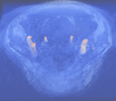
Acquisition parameters:
- FOV 180mm x 180mm, imaging matrix 512 x 512,
- 3 slabs resulting in 51 slices, slice thickness 0.5mm,
- TE=3.76 ms, TR=23ms, flip angle 25°,
- MAGNETOM Verio, Siemens Healthcare, 3T
Data Format:
- VTK STRUCTURED_POINTS float
- dimensions: 512 x 512 x 51
- Name: caroXXX_TOF.vtk (XXX stands for the dataset number)
Manual segmentations

- Separately for the right and left CCA/ICE/ECA
- VTK STRUCTURED_POINTS float
- Origin with regard to the whole dataset
- Tag to differentiate the region (0 = CCA, 1 = bifurcation, 2= ICA/ECA)
- Files:
- caroXXX_right_GS_YY.vtk
- caroXXX_left_GS_YY.vtk
- caroXXX_config.m (origin and tag for right/left segmentation)
Contact
For questions or comments please feel free to contact  Jana Hutter and refer to the publication:
[Hutter et al. 2012] Jana Hutter, Hannes G. Hofmann, Robert Grimm, Andreas Greiser, Marc Saake, Joachim Hornegger, Arnd Dörfler, Peter Schmitt: "Prior-based Automatic Segmentation of the Carotid Artery Lumen in TOF MRA", MICCAI 2012, LNCS (to appear)