
Contact
 +49 9131 9189774
 +49 9131 9189772

Address
Chair of Computer Science 5 (Pattern Recognition)
Henkestr. 91
91052 Erlangen
Germany
Powered by

Dr.-Ing. Michael Balda M. Sc.
Alumnus of the Pattern Recognition Lab of the Friedrich-Alexander-Universität Erlangen-Nürnberg
Quantitative CT
Quantitative Computed Tomography (QCT) generates quantitative values from datasets of reconstructed CT attenuation measurements. These values should only depend on the properties of the underlying materials.
Some methods of QCT yield quantitative values that represent different physical quantities: Rho-Z-projection for instance yields density and effective atomic number of the underlying material mixture.
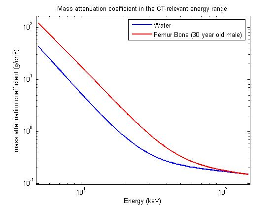
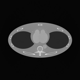
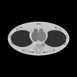
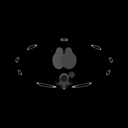
Basis material decomposition yields the relative densities of selected basis materials so that a mixture of these materials models the physical attenuation of the underlying material. For common body tissues an appropriate set of basis materials may consist of water and bone. Image 1 shows the mass attenuation coefficients of water and femur bone in cm²/g. Image 2 shows a thorax phantom and image 3 shows the according density images.
 |
 |
 |
 |
Data Acquisition
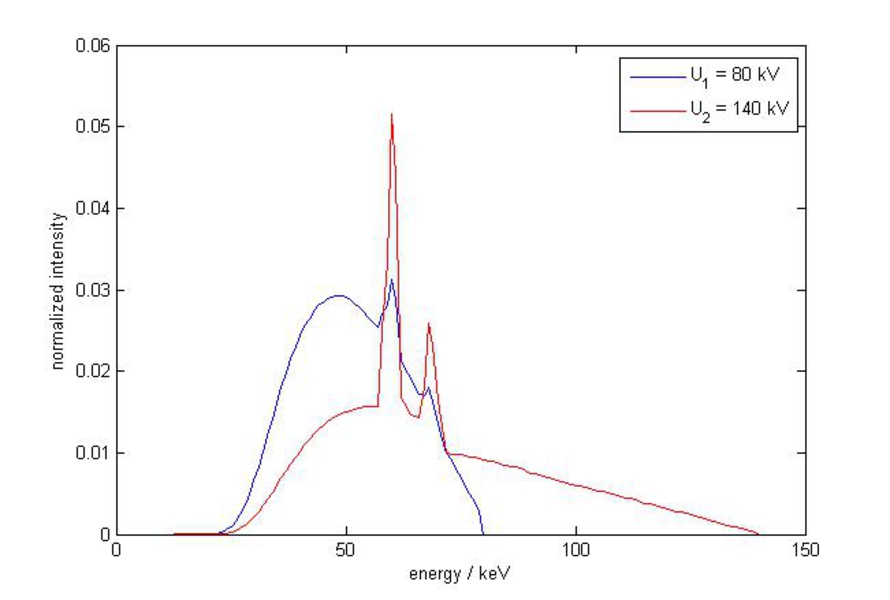
Monoenergetic radiation or multiple measurements at different system weightings are required for Quantitative CT. As common CT X-ray tubes usually produce polyenergetic radiation, the latter apporach is has to be chosen in medical CT.
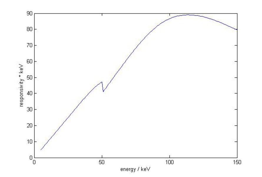
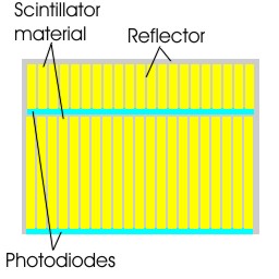
The system weighting consists of the spectrum of the incoming radiation and the detector responsivity, so changing one of these or both properties results in a different system weighting (see Img. 4). A strong separation between the system weightings is desired as transforms to quantitative values get more robust and therefore less vulnerable to noise. Different spectra can be produced by switching the tube voltage between readings (kVp-switching) or using two distinct measurement systems in one gantry (Dual-Source CT). The latter approach uses two X-ray tubes at different acceleration voltages possibly with different pre-filters. This way a strong separation between the spectra can be achieved and an individual tube current can be selected for each X-ray tube but the measurement is impaired by cross-scatter from the other measurement system. As Dual-Source CT also requires two detectors it is also possible to use different detector responsivities. Sandwich detectors (see Img. 5) can produce two or more measurements at different responsitivities in one reading by using a stack of scintillation detectors above each other. This has the advantage that the readings are prefectly aligned with respect to the gantry rotation angle, the dose usage, however, is inferior and the different weightings show a huge overlap. Unlike integrating detectors like he commonly used scintillators, Counting detectors are able to distinguish between incoming photon energies. Multiple photon counts different energy thresholds can be retrieved from single measurements.
 |
 |
 |