Contact
 +49 9131 85 28982
 +49 9131 85 27270

Address
Chair of Computer Science 5 (Pattern Recognition)
Martensstraße 3
91058 Erlangen
Germany
Powered by


Dr.-Ing. Mathias Unberath
Researcher in the Computed Tomography: Algebraic Reconstruction and Motion (CTARM) group at the Pattern Recognition Lab of the Friedrich-Alexander-Universität Erlangen-Nürnberg
I am a member of the  Graduate School in Advanced Optical Technologies at the Friedrich-Alexander Universität Erlangen-Nürnberg.
-
Various types of heart disease change left ventricular (LV) rotation. Measures of LV rotation are correlated with early onset symptoms of heart disease such as subclinical aortic stenosis and diastolic dysfunction. Noninvasive assessment of LV torsion, however, is challenging with modalities other than tagged MRI, which oftentimes is contraindicated due to metal implants.
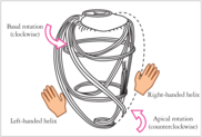
Coronary angiography is the imaging modality of choice for heart diseases. Virtually all clinical cases undergo this procedure at least once. In coronary angiography the heart arteries are selectively contrasted using a catheter that is inserted into the cardiac vessel system, making them clearly visible in projection X-ray imaging. As the coronary arteries are attached to the heart’s surface, they follow LV torsion.
The proposed research project aims at the development of a segmentation and reconstruction algorithm for 3D + t coronary angiograms in order to fully describe myocardial surface motion including torsion that is invisible in Computed Tomography and Magnetic Resonance Imaging. As the proposed project re-uses image data from standard imaging protocols, no additional radiation dose and discomfort is caused to the patient.
Articles in Conference ProceedingsProceedings of the Fully3D (13th International Meeting on Fully Three-Dimensional Image Reconstruction in Radiology and Nuclear Medicine), Newport, RI, 31.5.2015-4.6.2015, pp. 651-654, 2015 (BiBTeX, Who cited this?)
-
Interventional cardiac imaging is based on angiographic C-arm systems. In complex cardiac procedures
3D information can provide easier assessment and better guidance. Due to the low temporal resolution of such rotational angiography systems, 3D or even 4D (3D plus time) reconstruction of cardiac vasculature remains challenging, as motion artifacts corrupt image quality, reducing usability for diagnosis and guidance.
Recently, considerable progress has been made in the field of motion-compensated reconstruction of the heart chambers from C-arm CT data. Additionally, methods for automatic, interventional, quantitative left ventricular wall motion in 4D was proposed. Typically, the performance of such algorithms is evaluated using simulated data, as the ground truth is unknown in most of the clinical cases. For simulated data generation 3D and 4D numerical phantoms are forward projected using hardware-accelerated techniques such as ray casting.
Meaningful performance comparison of distinct motion-compensation techniques developed by different research groups requires use of projection data originating from the same phantom and geometry. While simulating different geometries is possible with virtually all simulation frameworks, using the same phantom can be challenging as most models are not publicly advertised. The 4D extended cardiac-torso (XCAT) phantom developed by Segars et al. constitutes an exception. Initially introduced in 1999, it is the most frequently used numerical cardiac phantom up to date. Since then, the spline-based phantom has been significantly advanced. The underlying 3D model is based on the Visible Human male and female anatomical data sets. The 3D model was then extended to 4D using deformations obtained from one tagged cardiac MRI data set and one respiratory-gated CT data set from normal male patient volunteers. As a consequence its clinical relevance can be doubted when changing clinical parameters, such as the ejection fraction, or when modeling pathologies. The XCAT phantom was developed with applications in SPECT and PET in mind. Due to the low spatial resolution of these modalities fewer anatomical details have to be modeled. For the evaluation of motion-compensated cardiac angiography reconstruction, however, the above properties of the XCAT are problematic as the goal of such algorithms is time resolved, volumetric reconstruction of pathologic hearts with the highest achievable resolution. A possible solution to aforementioned problems is the use of a statistical approach to shape modeling, as those models learn patterns of variability from a training set of correctly annotated shapes.
While the XCAT is publicly available against payment of a licensing fee, statistical shape models of the cardiac anatomy and dynamics are not yet disposable. We propose a highresolution, open-source 4D numerical phantom of the whole heart for the use in simulations of X-ray projection imaging. To the best of our knowledge, this is the first freely available numerical phantom of its kind.
Further information can be found on the project homepage of the
CONRAD Cardiac Model.
This project is in collaboration with the
Department of Radiology at Stanford University, Stanford, CA, USA.Articles in Conference ProceedingsProceedings of the ISBI (Biomedical Imaging, 2015 IEEE 12th International Symposium on), Brooklyn, NY, 16-19.4.2015, pp. 739-742, 2015 (BiBTeX, Who cited this?)
-
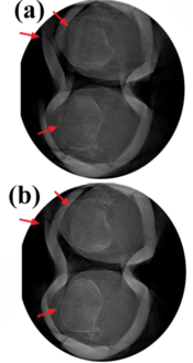
Recent advances in cone-beam Computed Tomography allow for the image acquisition of joints under weight-bearing conditions. However, images acquired under weight-bearing conditions are susceptible to severe motion artifacts because involuntary knee motion is common during data acquisition. Previous studies required the use of fiducial markers, which were used in four different correction methods in two dimensions (2D projection shifting, 2D projection warping) or three dimensions (3D image warping and 3D iterative image warping) for involuntary motion compensation. The 3D methods worked best because they were able to more accurately describe the effects on the projections of involuntary lower body motion. Nevertheless, the more restricted 2D projection shifting approach also showed significant reduction of motion-related artifacts. To overcome the need for fiducial markers, we propose an image-based 2D projection-shifting algorithm.
This work is a first step towards automatic image-based compensation for involuntary motion in weight-bearing CT scanning of knees.
Image on the right: (a) before and (b) after image-based compensation.
This project is in collaboration with the Department of
Radiology at Stanford University, Stanford, CA, USA.
Articles in Conference ProceedingsProc. SPIE Medical Imaging 2015 (SPIE Medical Imaging 2015), Orlando, FL, 21-26.2.2015, vol. 9413, pp. 94130D-7, 2015 (BiBTeX, Who cited this?) -