
Contact


Address
Chair of Computer Science 5 (Pattern Recognition)
Germany
Powered by


Dipl.-Inf. Rüdiger Bock
Alumnus of the Pattern Recognition Lab of the Friedrich-Alexander-Universität Erlangen-Nürnberg
Statistical Deformation Model of the Optic Disk
Statistical deformation model of the optic disk
Investigative Ophthalmology & Visual Science (IOVS), 2010, 51, ARVO E-Abstract 1774 (Poster as PDF, ARVO E-Poster)
Quantitative glaucoma indices commonly rely on geometric parameters of the optic nerve head (ONH) such as disk area or cup volume.
However, the ONH is too complex structure to be accurately analyzed by geometric morphometry because of:
- Sparse sampling of ONH
- Lack of mathematically derived and biologically interpretable ONH descriptions [2]
We provide dense descriptions of the ONH variability in glaucomatous cases presented by tissue deformations.
Erlangen Glaucoma Registry
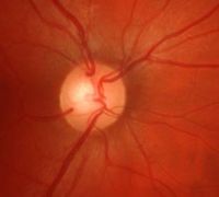
- Color fundus photographs (Kowa non-myd, FOV 22.5°, ONH centered)
- Gold standard for glaucoma diagnosis is present
- Diagnosis by an experienced ophthalmologist
- Complete ophthalmological examination
(ophthalmoscopy, visual field test, IOP, FDT, HRT II)
Dataset characteristics
- Mean age 55.4 ± 10.9 years
- ONH area 2.2 ± 0.5 mm2 (Macro ONH not included)
- 149 glaucoma cases Ig (FDT test time 67.4 ± 35.6 s)
- 246 controls Ic
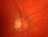
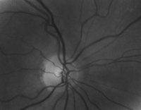
Fundus image preprocessing
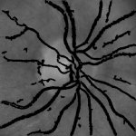
Normalized optic nerve head images by eliminating disease independent variations such as
- illumination inhomogeneities
- ONH normalization
- vessel structures.
Non rigid image registration
Characterization of inter subject ONH variability by dense deformation fields calculated between one reference image and image samples
- Variational multilevel approach D(R(U), T) + s * S(U) →U min
- Distance measure D: Sum of squared differences
- Smoother S: Diffusion regularizer
Deformation based reference ONH
ONH reference image R that is characterized by the minimal average residual deformation over a sample set I is calculated by an iterative procedure:
- Reference R: Selection of initial estimate as the average of sample set I
- Calculation of deformation fields between samples and current reference
- Average all deformation fields to average deformation field
- Deformation of reference by average deformation field
Statistical deformation model (SDM) of ONH
to provide a mathematically derived and biologically interpretable description of ONH variability in glaucomatous optic atrophy.
- PCA model on deformations of
- control samples Ic
- glaucomatous cases Ig
- Supervised attribute selection on modes
→ Identification of modes most discriminating between controls and glaucomatous
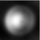
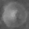
Deformation based reference ONH
Comparison of pixel wise averaging (left) and deformation based averaging
(right) of ONH reflectance images of controls.
Pixel wise averages are too smooth to indentify local structures. Deformation based averaging clearly shows the naturally shaped appearance of the cup (central bright spot).
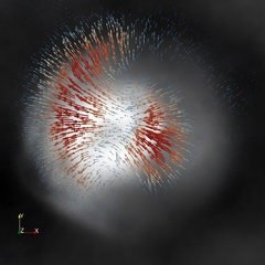
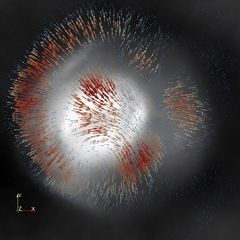
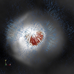
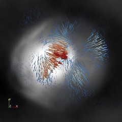
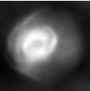
Statistical Deformation Model of ONH in glaucoma
First four PCA modes most discriminating between control and glaucoma
providing a mathematically derived and biologically interpretable description of ONH deformations: (first row) Simultaneous deformation of the cup and rim, (second row) local extension of the cup (red: high magnitude, blue: low magnitude of deformation vector).
- Discriminative modes identify patterns of variation
- Intuitive visualization of ONH deformation (Figure 4)
- Mathematically derived influence of rim and cup
→ Biologically interpretable descriptions of ONH deformation in case of glaucoma
Statistical deformation modelling of ONH provides
- intuitive representation of ONH variability
- main modes of glaucomatous deformations
- identification of abnormal deformations.
Deformation based morphometry of ONH shows a potential to be a future technique to gain novel insights into glaucomatous changes of the ONH.