
Contact

 +49-9131-85-27775
 +49-9131-85-27270
Secretary
| Monday | 8:00 - 12:15 |
| Tuesday | 8:00 - 16:45 |
| Wednesday | 8:00 - 16:45 |
| Thursday | 8:00 - 16:45 |
| Friday | 8:00 - 12:15 |
Address
Lehrstuhl für Informatik 5 (Mustererkennung)
Martensstr. 3
91058 Erlangen
Germany
Powered by

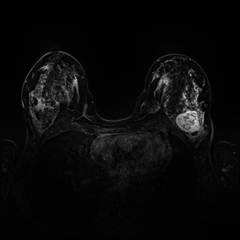
Breast Tumor Lesions Segmentation
There are several imaging techniques used for screening, diagnostics and tumor progression analysis. The most common approach for screening is conventional mammography. However, mammograms present certain limitations, First, there is a possibility to wrongly identify lesions as tumor cancer which occurs when radiologists decide mammograms are abnormal but no cancer is actually present. This also resulted in the further testing process such as ultrasound and biopsy. Second, overdiagnosis and overtreatment might happen when mammograms find cancers Third, the most important limitation on x-rays mammograms comes from the fact that Fatty tissue appears darker on a mammogram, whereas fibroglandular tissue appears as white areas. The reason behind this is that fibroglandular tissue and tumors have the similar density that is why tumors are harder to detect in women with denser breasts.
On another hand, the breast MRI procedure presents several advantages over the Traditional x-ray mammograms. Breast MRI obtains 3D images with the particular advantage of providing a strong contrast between fatty tissue and fibroglandular tissue, ideal for calculating the breast density factor. These detailed pictures obtained on the exam can show the difference between normal and diseased tissue. One of the most significant advantages of an MRI exam is that the patient does not receive any marrying radiation such as X and Gamma ray.
Group members in this project:  Sulaiman Vesal
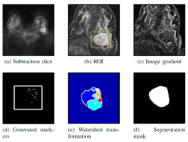
An accurate and precise segmentation of the breast lesion in a Computer Aided Detection (CAD) system is a crucial step in evaluating tumor volume and in the quantification of tumor characteristics. However, this is a challenging task, since breast lesions have sophisticated shape, topological structure, and variation in the intensity distribution. In this paper, we proposed a novel Marker-controlled Watershed method which uses brightest pixels as markers to overcome this challenge and accurately segment the breast MRI lesions through predetermined Region of Interest (ROI) by an expert. The proposed approach was evaluated on 106 lesion cases which include 59 malignant and 47 benign. The segmentation results compared with ground truth through Jaccard and Dice Coefficient metrics. The results illustrate that the proposed method significantly improve segmentation accuracy and it will help further in improving classification performance between malignant and radiologically suspicious benign lesions.