Dr.-Ing. Tanja Kurzendorfer
Alumnus of the Pattern Recognition Lab of the Friedrich-Alexander-Universität Erlangen-Nürnberg
-
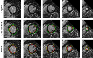
The anatomy of the left ventricle (LV) along with a detection of scar tissue in the myocardial muscle provides important information for the treatment of heart failure. Heart failure often arises from ventricular dyssynchrony, when left and right ventricles are not contracting at the same time. This dyssynchrony can be treated with the implantation of a bi-ventricular pacemaker in cardiac resynchronization therapy (CRT). However, the presence of scar tissue hinders the electrical signal coming from the CRT device, which negatively affects the success of the treatment. Integrating the knowledge of the location of scar tissue to treatment planning could improve the efficacy of CRT.
Commonly, Late-Gadolinium-Enhanced (LGE) MR imaging is performed to visualize both scar tissue and the anatomy of the heart. The use of such images for therapeutic procedures currently lacks automatic segmentation approaches. Manual segmentation methods are available, but their use is time consuming and tedious. Therefore, it would be desirable if the segmentation of the myocardium and the scar classification were completely automatic and independent of any manual interaction.
The current research focus is on fully automatic LV anatomy and scar segmentation from LGE-MRI.
 Articles in Conference ProceedingsBildverarbeitung für die Medizin 2017, Heidelberg, 12-14.03, pp. 11-16, 2017 (BiBTeX, Who cited this?)Proceedings of the 2017 IEEE International Symposium on Biomedical Imaging: From Nano to Macro (2017 IEEE 14th International Symposium on Biomedical Imaging (ISBI)), Melbourne, 18-21.04, pp. 831-834, 2017 (BiBTeX, Who cited this?)Book of Abstracts (11th Interventional MRI Symposium), Baltimore, 7-8.10, pp. P-03, 2016 (BiBTeX, Who cited this?)32nd Annual Scientific Meeting of the ESMRMB (ESMRMB 2015), Edinburgh, 1-3.10, pp. 318-319, 2015 (BiBTeX, Who cited this?)

 +49 9131 85 27874
 +49 9131 85 27270

