Contact
 +49 9131 85 27891
 +49 9131 85 27270

Address
Chair of Computer Science 5 (Pattern Recognition)
Martensstr. 3
91058 Erlangen
Germany
Powered by


Bastian Bier M. Sc.
Alumnus of the Pattern Recognition Lab of the Friedrich-Alexander-Universität Erlangen-Nürnberg
-
Detection results for different viewing angles. 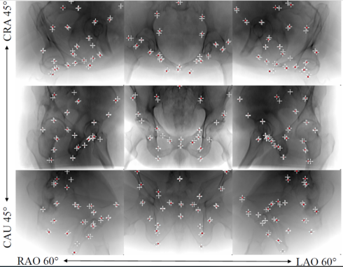
X-ray image guidance enables percutaneous alternatives to complex procedures. Unfortunately, the indirect view onto the anatomy in addition to projective simpli cation substantially increase the taskload for the surgeon. Additional 3D information such as knowledge of anatomical landmarks can bene t surgical decision making in complicated scenarios. Automatic detection of these landmarks in transmission imaging is challenging since image-domain features characteristic to a certain landmark change substantially depending on the viewing direction. Consequently and to the best of our knowledge, the above problem has not yet been addressed. In this work, we present a method to automatically detect anatomical landmarks in X-ray images independent of the viewing direction. To this end, a sequential prediction framework based on convolutional layers is trained on synthetically generated data of the pelvic anatomy to predict 23 landmarks in single X-ray images. View independence is contingent on training conditions and, here, is achieved on a spherical segment covering 120°/90° in LAO/RAO and CRAN/CAUD, respectively, centered around AP. On synthetic data, the proposed approach achieves a mean prediction error of 5.6 +- 4.5 mm. We demonstrate that the proposed network is immediately applicable to clinically acquired data of the pelvis. In particular, we show that our intra-operative landmark detection together with pre-operative CT enables X-ray pose estimation which, ultimately, bene ts initialization of image-based 2D/3D registration.
Articles in Conference ProceedingsMedical Image Computing and Computer-Assisted Intervention (MICCAI 2018), Granada, Spain, 17. - 19.09.2018, pp. 55-63, 2018 (BiBTeX, Who cited this?)
-
(Top) Geometry of the acquisition.
(Bottom) Uncorrected and corrected reconstruction.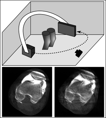
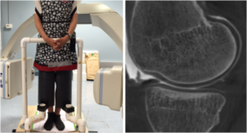
Cone-beam C-arm CT systems allow to scan patients in weight-bearing positions to assess knee cartilage health under more realistic conditions. Involuntary patient motion during the acquisition results in motion artifacts in the reconstructions. The current motion estimation method is based on fiducial markers. They can be tracked with a high spatial accuracy in the projection images, but only deliver sparse information. Further, placement of the markers on the patient's leg is time consuming and tedious.
Instead of relying on a few well defined points, we seek to establish correspondences on dense surface data to estimate 3D displacements. In this feasibility study, motion corrupted X-ray projections and surface data are simulated. We investigate motion estimation by registration of the surface information.
The proposed approach is compared to a motion free, an uncompensated, and a state-of-the-art marker-based reconstruction using the SSIM. The proposed approach yields motion estimation accuracy and image quality close to the current state-of-the-art, reducing the motion artifacts in the reconstructions remarkably. The Structural Similarity improved from 0.887 to 0.975 from the uncorrected images using the proposed approach. The results are promising and encourage future work aiming at facilitating its practical applicability.
Journal ArticlesRange Imaging for Motion Compensation in C-Arm Cone-Beam CT of Knees under Weight-Bearing ConditionsJournal of Imaging, vol. 4, no. 1, pp. 1-16, 2018 (BiBTeX, Who cited this?)Articles in Conference ProceedingsMedical Image Understanding and Analysis (21st Annual Conference, MIUA 2017), Edinburgh, 11.07.2017-13.07.2017, pp. 561, 2017 (BiBTeX, Who cited this?)2017 IEEE Nuclear Science Symposium and Medical Imaging Conference Record (NSS/MIC) (IEEE Nuclear Science Symposium and Medical Imaging Conference (NSS/MIC)), Atlanta, 21.10-28.10.2017, pp. tbd, 2017 (BiBTeX, Who cited this?)
-
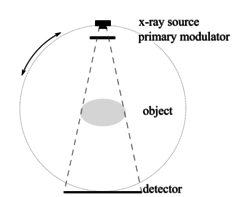
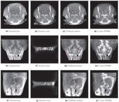
Cone beam computed tomography (CBCT) suffers from a large amount of scatter, resulting in severe scatter artifacts in the reconstructions. Recently, a new scatter correction approach, called improved primary modulator scatter estimation (iPMSE), was introduced. That approach utilizes a primary modulator that is inserted between the X‐ray source and the object. This modulation enables estimation of the scatter in the projection domain by optimizing an objective function with respect to the scatter estimate. Up to now the approach has not been implemented on a clinical angiography C‐arm CT system. In our work, the iPMSE method is transferred to a clinical C‐arm CBCT. Additional processing steps are added in order to compensate for the C‐arm scanner motion and the automatic X‐ray tube current modulation. These challenges were overcome by establishing a reference modulator database and a block‐matching algorithm. Experiments with phantom and experimental in vivo data were performed to evaluate the method. We show that scatter correction using primary modulation is possible on a clinical C‐arm CBCT. Scatter artifacts in the reconstructions are reduced with the newly extended method. Compared to a scan with a narrow collimation, our approach showed superior results with an improvement of the contrast and the contrast‐to‐noise ratio for the phantom experiments. In vivo data are evaluated by comparing the results with a scan with a narrow collimation and with a constant scatter correction approach. Scatter correction using primary modulation is possible on a clinical CBCT by compensating for the scanner motion and the tube current modulation. Scatter artifacts could be reduced in the reconstructions of phantom scans and in experimental in vivo data.
Journal ArticlesMedical Physics, vol. 44, no. 9, pp. e125-e137, 2017 (BiBTeX, Who cited this?)Articles in Conference ProceedingsProceedings of the 4th International Conference on Image Formation in X-ray Computed Tomography (CT-Meeting 2016), Bamberg, 18.07.2016-22.07.2016, pp. 383-386, 2016 (BiBTeX, Who cited this?)
 |
Osteoarthritis (OA) is the leading cause of functional decline and disability in aging populations. The causes and progression of OA, particularly in the early stages, remain poorly understood. Current OA imaging measures are insensitive to early changes, or are logistically challenging and limited by expense and long scan times.
The overarching goal of this project is to develop a novel weight-bearing computed tomography (CT) imaging method to expand our understanding of the mechanical stresses that affect cartilage and meniscus health and to provide a quantitative measure of knee joint health.
Therefore, patients are scanned in standing upright or squatting position. In contrast to acquisitions in supine position, patients are more likely to show involuntary motion which results in motion artifacts in the reconstructions decreasing the diagnostic image quality. Focus of current research is the development of novel motion correction approaches using surface cameras.
More informations about the project can be found  here.
here.
The project is in collaboration with the Department of Radiology, Stanford University, Stanford, CA, USA.
I am a member of the research training group "Heterogeneous Image Systems" http://hbs.fau.de/.